
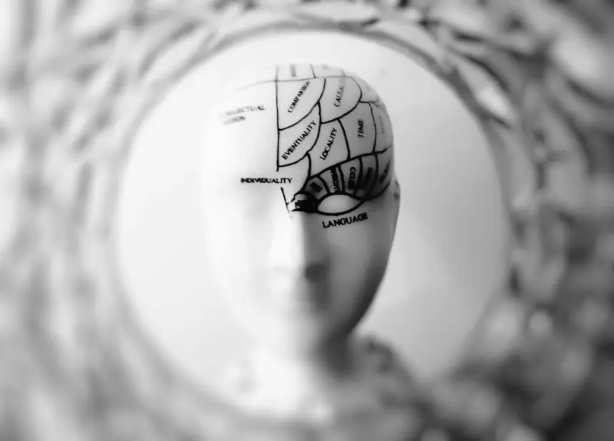
The brain is the most complex organ in the human body. It defines our personality, guides our movements, and interprets the world around us. Every thought, emotion, and memory begins in this intricate network of billions of neurons working in harmony.
- The Brain’s Basic Architecture
- 2. The Cerebellum: Balance and Coordination
- 3. The Brainstem: The Body’s Life Support System
- 4. The Limbic System: Emotion and Memory
- 5. The Ventricular System and Cerebrospinal Fluid
- 6. The Blood-Brain Barrier
- How Brain Regions Work Together
- The Nervous System Connection
- Plasticity: The Brain’s Remarkable Ability to Adapt
- Why Understanding Anatomy Matters in Brain Injury Care
- The Big Picture: An Organ of Infinite Complexity
Understanding how the brain is structured — and what each part does — is essential for grasping how brain injuries affect people differently. The location and severity of damage determine whether someone struggles with memory, movement, speech, or even emotional control.
The Brain’s Basic Architecture
Protected by the skull and cushioned by fluid, the brain weighs about three pounds, yet it consumes nearly 20 percent of the body’s oxygen and energy. It is divided into major regions, each performing specialized but interconnected roles.
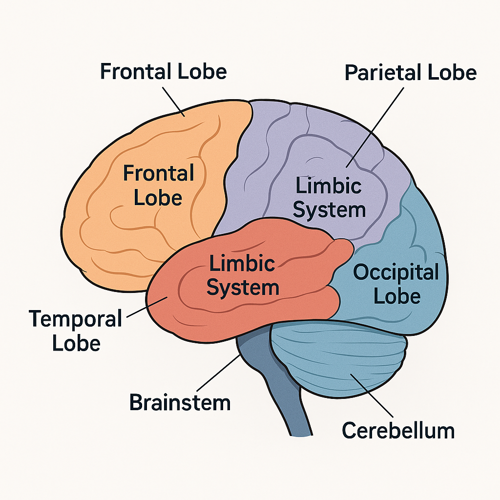
1. The Cerebrum
The cerebrum is the largest and most recognizable part of the brain. It’s divided into left and right hemispheres, connected by a thick band of nerve fibers called the corpus callosum.
Each hemisphere controls the opposite side of the body — the left hemisphere governs the right side, and vice versa. Within the cerebrum are four lobes, each responsible for distinct functions:
- Frontal lobe: The center of reasoning, judgment, planning, and voluntary movement. It also plays a key role in impulse control and emotional regulation. Damage here can cause behavioral changes or difficulty with decision-making.
- Parietal lobe: Processes sensory input such as touch, pressure, and spatial awareness. Injury may lead to difficulty recognizing objects or judging distances.
- Temporal lobe: Houses the auditory cortex and memory centers. It helps with hearing, language comprehension, and emotional memory.
- Occipital lobe: Dedicated to vision. Even minor damage here can lead to partial or total loss of sight.
Together, these lobes make the cerebrum the command center for thought, creativity, and behavior.
2. The Cerebellum: Balance and Coordination
Beneath the cerebrum sits the cerebellum — smaller, but densely packed with neurons. It fine-tunes voluntary movements, balance, and coordination.
When the cerebellum is injured, people may struggle with posture, walking, or precise hand movements. They might appear clumsy or have difficulty performing tasks that once felt automatic.
While it doesn’t initiate movement, the cerebellum ensures that every step, reach, and gesture is smooth and efficient.
3. The Brainstem: The Body’s Life Support System
Connecting the brain to the spinal cord, the brainstem regulates the functions we rarely think about but cannot live without — breathing, heart rate, and sleep-wake cycles. It has three main parts:
- Midbrain: Coordinates eye movement and visual or auditory reflexes.
- Pons: Acts as a bridge between the cerebrum and cerebellum; helps control breathing and facial sensations.
- Medulla oblongata: Regulates vital functions such as heartbeat, swallowing, and blood pressure.
Damage to the brainstem can be catastrophic, as even small injuries may interrupt the body’s automatic life-support systems.
4. The Limbic System: Emotion and Memory
Deep within the cerebrum lies the limbic system — a group of interconnected structures that link emotion, memory, and behavior. It includes the hippocampus, amygdala, and parts of the thalamus and hypothalamus.
- Hippocampus: Essential for forming new memories and spatial navigation.
- Amygdala: Regulates fear, anger, and emotional learning.
- Hypothalamus: Maintains homeostasis — controlling hunger, thirst, temperature, and hormone release.
When brain injury disrupts the limbic system, individuals may experience emotional instability, memory loss, or exaggerated stress responses.
5. The Ventricular System and Cerebrospinal Fluid
The brain contains a series of interconnected cavities called ventricles, filled with cerebrospinal fluid (CSF). This clear liquid cushions the brain from impact, delivers nutrients, and removes waste.
Disruption of normal CSF flow, such as in hydrocephalus, can cause dangerous increases in pressure inside the skull—something that must be treated promptly to prevent permanent damage.
6. The Blood-Brain Barrier
The brain is protected by a selective filter called the blood-brain barrier. This barrier allows nutrients like glucose and oxygen to pass through while blocking toxins and pathogens.
While crucial for protection, this barrier can also complicate treatment, as many medications cannot easily cross it. This is one reason why brain injuries and neurological diseases are often difficult to manage pharmacologically.
How Brain Regions Work Together
No part of the brain works alone. For example, speaking a simple sentence engages multiple regions: the frontal lobe plans and executes movement, the temporal lobe interprets meaning, and the cerebellum refines the coordination of speech muscles.
Similarly, memory, emotion, and movement are tightly linked. This interconnectedness explains why a single injury can have far-reaching effects — why damage to the frontal lobe may affect motivation, or why a cerebellar injury can impair cognitive speed.
The Nervous System Connection
The brain is part of a larger communication network: the central nervous system (CNS) and peripheral nervous system (PNS).
The CNS consists of the brain and spinal cord, while the PNS carries messages to and from the body through sensory and motor nerves.
When brain injury occurs, the flow of these messages is disrupted. Depending on which neural pathways are affected, a person may experience paralysis, sensory loss, or changes in reflexes.
Plasticity: The Brain’s Remarkable Ability to Adapt
One of the most hopeful discoveries in neuroscience is neuroplasticity—the brain’s ability to reorganize itself after injury. When one area is damaged, nearby regions can sometimes take over its functions. This adaptability underlies the entire field of brain injury rehabilitation.
Therapies like cognitive rehabilitation, physical therapy, and speech-language therapy harness neuroplasticity by stimulating neural circuits to re-learn lost skills.
However, plasticity has limits. Recovery depends on many factors, including age, overall health, the type of injury, and early intervention. The more actively and consistently the brain is challenged, the greater the chance of regaining function.
Why Understanding Anatomy Matters in Brain Injury Care
Clinicians and therapists study brain anatomy to predict how a patient’s symptoms may evolve. A person with frontal lobe damage may require behavioral and decision-making support; someone with temporal lobe damage might need help with language or memory cues.
Families can also benefit from understanding anatomy — it turns confusion into clarity, helping them interpret why their loved one behaves or feels differently after injury. Knowledge empowers caregivers to communicate effectively with medical teams and recognize early signs of complications.
The Big Picture: An Organ of Infinite Complexity
The human brain is more than a collection of lobes and neurons — it’s a living network that defines consciousness itself. Every heartbeat sends oxygen to sustain it; every experience reshapes its connections.
When injury strikes, understanding this organ’s architecture reveals both its fragility and resilience. The same pathways that allow trauma to cause damage also enable healing, adaptation, and growth.
In learning how the brain works, we come to appreciate not just its complexity, but its extraordinary capacity to recover, redefine, and relearn.