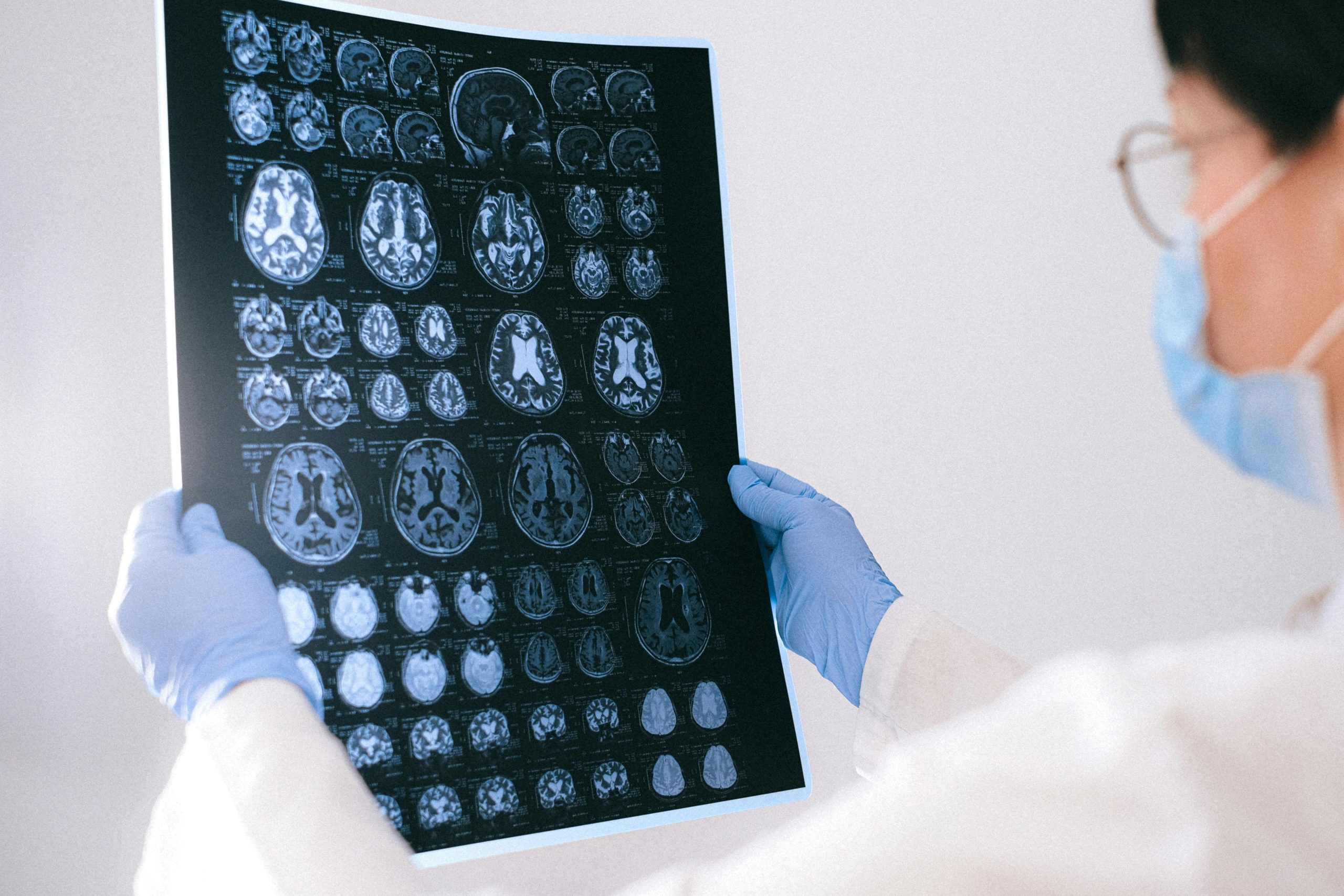
After a brain injury, one of the first things that happens is a scan, usually a CT, and sometimes an MRI later on. The words get used quickly, results are discussed in medical language, and it can be hard to know what any of it means. Understanding what these scans are, and what they can and cannot show, makes the conversations with the care team a little easier to follow.
This is a plain-language guide to the two main brain scans and what to expect from them. It is here for orientation, not to interpret a particular result. What a scan shows for your loved one, and what it means, is a question for the care team, who can read it alongside everything else they know.
Key takeaways
- A CT scan is fast and usually done first, because it quickly shows bleeding, fractures, and swelling.
- An MRI gives more detail and can reveal subtler injury, but takes longer and is usually done later.
- A normal scan does not always mean no injury. Some damage is too fine to show up.
- A scan is one piece of the picture, read alongside the exam and how a person responds.
Why scans are used
After a head injury, the care team needs to see what is happening inside the skull, and they cannot do that from the outside. Scans let them look for bleeding, swelling, fractures, and other changes, so they can decide what treatment is needed and how urgently. In the early stage, imaging can genuinely be life-saving, because it reveals problems such as a bleed that may need surgery. Later, scans help the team follow how things are changing over time.
The CT scan
A CT scan, short for computed tomography, uses X-rays to build detailed cross-sectional pictures of the brain and skull. It is almost always the first scan after a serious head injury, for one simple reason: it is fast, widely available, and very good at spotting the things that matter most in an emergency. A head CT often takes only minutes, and the scan itself is painless. A CT is especially good at showing:
- ›Bleeding in or around the brain, such as a hematoma.
- ›Skull fractures.
- ›Swelling and signs of raised pressure inside the skull.
During the scan, the person lies on a table that slides through a ring-shaped opening. It is quicker and more open than an MRI, which is part of why it suits someone who is seriously unwell. It does involve a small dose of radiation, which is considered low for a single scan and is weighed against the clear benefit of seeing inside quickly. Sometimes a contrast dye is given through a vein to show certain things more clearly.
The MRI scan
An MRI, short for magnetic resonance imaging, uses strong magnets and radio waves rather than X-rays, so it involves no radiation. It produces even more detailed pictures than a CT and can reveal subtler injuries that a CT may miss, which is why it is often used a little later, once someone is stable, to understand an injury more fully. An MRI can show:
- ›Small bruises and areas of damage that a CT cannot pick up.
- ›Diffuse axonal injury, the widespread stretching and tearing of nerve fibers, which is notoriously hard to see on an early CT. Our guide on diffuse axonal injury explains this.
- ›Detail about soft tissue and the structure of the brain.
The trade-off is that an MRI takes longer, often much longer than a CT, and the machine is a narrower tunnel and quite loud, with knocking and buzzing sounds. The person needs to stay still, which is one reason it is not always the first choice for someone who is critically unwell or cannot lie still. Because it uses powerful magnets, metal is a safety issue, so the team will ask carefully about any implants or metal in the body.
Why the team chooses one or the other
It is not that one scan is better and the other worse. They are different tools for different moments. A CT is the workhorse of the emergency, fast and excellent at finding urgent problems like bleeding. An MRI is the detailed follow-up, better at revealing subtle or widespread injury once there is time and the person is stable enough. Many people with a serious injury will have a CT early and one or more MRIs later, and often repeated CT scans in between, so the team can watch for changes such as new bleeding or swelling. Seeing your loved one sent for scan after scan can be worrying, but it usually means the team is keeping a close and careful eye on things.
Why a “normal” scan does not always mean no injury
This is one of the most important and most confusing things for families. A scan can look close to normal even when there is a real and significant brain injury. This is especially true for diffuse axonal injury, where the damage is microscopic and spread out rather than a single obvious mark. In fact, a large share of these injuries do not show clearly on an early CT at all. It does not mean the injury is not real, and it does not mean the doctors have missed something. It means some kinds of damage are simply too fine to appear on a picture, which is why the team never relies on scans alone.
What to expect if your loved one needs a scan
If your loved one is having a scan, a few things may help you feel steadier. A CT is quick and painless, over in minutes. An MRI takes longer and is noisy, and they will usually be given earplugs or headphones. For both, they need to lie still, and for someone confused or agitated, the team may use gentle sedation to help. You often cannot be in the room during the scan itself, because of the radiation or the magnet, but the staff are watching the whole time and you can usually wait close by. Afterward, a radiologist, a doctor who specializes in reading scans, reviews the images and shares the findings with the care team, who will explain them to you. It is completely fair to ask when results will be ready and who will go through them with you.
Does every brain injury need a scan?
Not always. For a milder injury or a concussion, doctors use guidelines and their judgment to decide whether a scan is needed at all, because a scan is not always helpful and carries its own small considerations. If a scan is not ordered, it is not the team being dismissive. It often means they do not expect it to change the care, and that they are watching the person instead. If you are ever unsure why a scan is or is not being done, it is always reasonable to ask.
Common questions
Why did they do a CT and not an MRI?
A CT is faster, more widely available, and very good at quickly finding urgent problems like bleeding, fractures, and swelling, which makes it the natural first scan in an emergency. An MRI gives more detail but takes longer, so it is often used later, once someone is stable, to understand the injury more fully.
The scan looked normal, so why is my loved one so unwell?
Some brain injuries, especially diffuse axonal injury, are microscopic and do not show clearly on scans, particularly an early CT. A near-normal scan does not mean there is no injury. The team relies on the whole picture, including the exam and how the person responds, not the scan alone.
Is the radiation from a CT scan dangerous?
The radiation dose from a single head CT is considered low, and the benefit of seeing inside the skull quickly usually far outweighs it. Doctors are mindful of repeated scans and weigh each one. An MRI, by contrast, uses magnets and radio waves and involves no radiation at all.
Why does my loved one keep having repeated scans?
After a serious injury, the brain can keep changing, with swelling or new bleeding developing over hours and days. Repeated scans let the team catch these changes early and respond. It can be unsettling to watch, but it usually reflects careful monitoring rather than something going wrong.